Cancer remains one of the biggest health challenges of our time, claiming nearly 10 million lives each year. With global cases expected to rise in the coming decades, the need for better solutions is urgent.
What if technology could help us detect cancer earlier, treat it more effectively, and even predict its progression before symptoms appear?
Artificial intelligence (AI) and machine learning (ML) have already transformed industries like finance and entertainment, and now they’re making a profound impact on healthcare, especially in cancer research.
Using vast amounts of medical data, AI is helping researchers and clinicians tackle some of the toughest challenges in oncology. From spotting tumors in medical images faster than the human eye to utilizing medical image generation for more accurate diagnostics, AI and ML are reshaping cancer care, paving the way for earlier detection, more precise treatment, and better outcomes.
In this blog, we’ll explore how AI is driving progress in cancer research, from enhancing imaging techniques like medical image generation to accelerating the discovery of new drugs.
The Role of Machine Learning in Cancer Research
Machine learning (ML) is a branch of artificial intelligence that enables computer systems to learn from data and make predictions or decisions without being explicitly programmed for every task.
In cancer research, ML algorithms analyze large volumes of clinical, imaging, and genomic data to identify patterns and make accurate predictions. This ability is essential for improving early detection, diagnosis, and treatment outcomes.
ML models are beneficial in oncology because they can process complex datasets, such as medical images, pathology reports, or patient histories, more quickly and consistently than humans. As cancer diagnostics and treatments increasingly rely on large-scale data, machine learning (ML) provides a scalable and precise method for supporting clinical decisions.
Machine Learning Methods in Cancer Diagnostics and Treatment
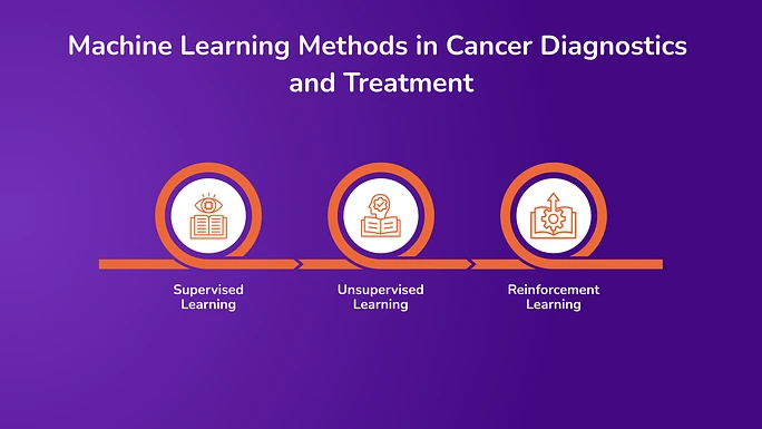
Below is an overview of the key machine learning methods that are playing a significant role in cancer diagnostics and treatment.
1. Supervised Learning
Supervised learning is the most commonly used machine learning (ML) method in cancer diagnostics. In this method, the algorithm is trained on a labeled dataset, meaning that each example in the dataset includes input data and the correct output (e.g., an image of a tumor and its known classification as benign or malignant).
Once trained, the model can predict the label for new, unseen data. In oncology, supervised learning is used in:
- Tumor detection in mammograms and CT scans.
- Classifying skin lesions as cancerous or non-cancerous.
- Predicting patient survival rates based on clinical features.
These models improve over time as more labeled data becomes available, helping to increase diagnostic accuracy and reduce false positives or negatives.
2. Unsupervised Learning
Unsupervised learning deals with datasets that have no predefined labels. The goal is to identify hidden patterns, clusters, or structures within the data. This technique is valuable when exploring large-scale biological data, such as gene expression profiles, where the relationships between variables are not clearly defined.
In cancer research, unsupervised learning is used for:
- Discovering new cancer subtypes by grouping patients with similar genetic or molecular profiles.
- Analyzing histopathology slides to identify uncommon cellular structures.
- Detecting anomalies in datasets that might indicate rare forms of cancer.
Unsupervised models help researchers uncover previously unknown insights without needing human-labeled data, which is often time-consuming and expensive to produce.
3. Reinforcement Learning
Reinforcement learning (RL) focuses on training models to make a sequence of decisions by rewarding desired behaviors and penalizing undesired ones. Over time, the model learns strategies that lead to better outcomes.
In oncology, RL is used in:
- Optimizing treatment schedules for radiation or chemotherapy.
- Modeling how cancer might evolve in response to therapy.
- Simulating personalized treatment plans to maximize effectiveness while minimizing side effects.
Reinforcement learning is beneficial in adaptive therapy, where treatment protocols adjust based on a patient’s ongoing response rather than following a fixed plan.
Clinical Applications of Machine Learning in Cancer Research
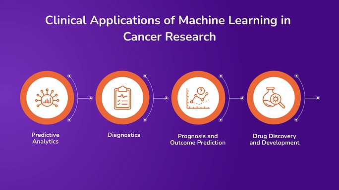
Machine learning is being applied across multiple stages of cancer research and care, from early detection to treatment planning and drug development. Below are the core clinical areas where machine learning (ML) has shown a measurable impact.
1. Predictive Analytics
Early Detection of High-Risk Patients Based on EHR Data
Machine learning models can analyze patterns in electronic health records (EHRs) to identify individuals at high risk of developing cancer. These models evaluate a combination of patient demographics, lab results, medical history, genetic data, and clinical notes to generate risk scores. This helps clinicians flag potential cases earlier, even before symptoms emerge, enabling earlier screenings and interventions.
Identifying Disease Progression Before Symptoms Arise
ML algorithms trained on longitudinal patient data can recognize subtle changes that precede clinical symptoms. For example, they can track how biomarker levels, imaging results, or vital signs change over time and correlate these trends with disease progression. This proactive approach supports timely adjustments in care, which can improve survival outcomes.
2. Diagnostics
ML Models Matching or Surpassing Dermatologists in Skin Cancer Classification
Deep learning models have been trained on thousands of dermatoscopic images to classify skin lesions. These models can differentiate between benign and malignant skin lesions with accuracy levels comparable to or better than those of experienced dermatologists. They are now being tested in real-world settings to assist in routine dermatological screenings, especially in areas with limited specialist availability.
Use in Colon Cancer Screening (e.g., Mismatch Repair Detection)
Machine learning is also being used to detect mismatch repair deficiency (dMMR), a marker for certain types of colorectal cancers. Traditional methods rely on complex and time-consuming lab tests, but machine learning (ML) can analyze genomic or histopathological data to identify dMMR more efficiently. This has implications for both diagnosis and the selection of targeted therapies, such as immunotherapy.
3. Prognosis and Outcome Prediction
Predicting Relapse, Metastasis, or Treatment Resistance
By analyzing patient history, treatment response data, and tumor characteristics, machine learning (ML) models can predict the likelihood of cancer relapse, spread to other organs, or resistance to specific therapies. These models help oncologists choose more aggressive monitoring for high-risk patients or modify treatment strategies in advance.
Risk Stratification for Better Clinical Decision-Making
ML tools help divide patients into low-, medium-, and high-risk groups based on various factors. This stratification supports more personalized care pathways, such as determining who may benefit from intensive therapy versus who may avoid overtreatment. It also informs clinical trial design by identifying patients who meet specific risk criteria.
4. Drug Discovery and Development
Identifying Potential Drug Candidates Faster and More Efficiently
Conventional drug discovery is a time-consuming and expensive process. Machine learning accelerates this by screening vast libraries of compounds and predicting their biological activity based on molecular structure. These predictions can narrow down the list of candidates for lab testing, saving time and resources.
Predicting Molecule Interactions and Simulating Clinical Trials
ML models can predict how new drug molecules will interact with proteins or cells related to cancer. In addition, simulations using machine learning (ML) can forecast how a drug will perform across diverse patient populations, reducing reliance on early-phase clinical trials. These insights help in prioritizing the most promising compounds for further development and approval.
Using Medical Image Generation to Overcome Data Limitations in Cancer Imaging with AI
One of the significant challenges in using machine learning (ML) for cancer imaging is the lack of high-quality, labeled images, especially for rare types of cancer. To make machine learning models work well, they require large amounts of data. However, collecting this data can be expensive, time-consuming, and limited by privacy rules.
Generative models can help solve this problem by creating synthetic, or fake, medical images that complement real datasets. There are two main types of generative models used:
Generative Adversarial Networks (GANs)
GANs use two networks that work together. One creates synthetic images, and the other checks if they look real or fake. Over time, the generator learns to make more realistic images. GANs have been used to:
- Create synthetic MRIs and CT scans.
- Simulate rare disease cases.
- Expand training datasets to reduce errors in predictions.
However, GANs can have some issues, such as unstable training and a limited variety of images they generate.
Diffusion Models
Diffusion models are a more stable and accurate way to generate images. They start with noise and gradually turn it into a clear picture through a series of steps. These models:
- Can create realistic 3D images, such as MRIs of brain tumors
- Works well with small datasets, which is helpful for rare cancers.
This will allow researchers to train models using fewer images by using text to guide the process, resulting in highly accurate and relevant images for clinical use.
These models are helping researchers create more diverse and realistic medical images, making it easier to train machine learning models for cancer detection and treatment.
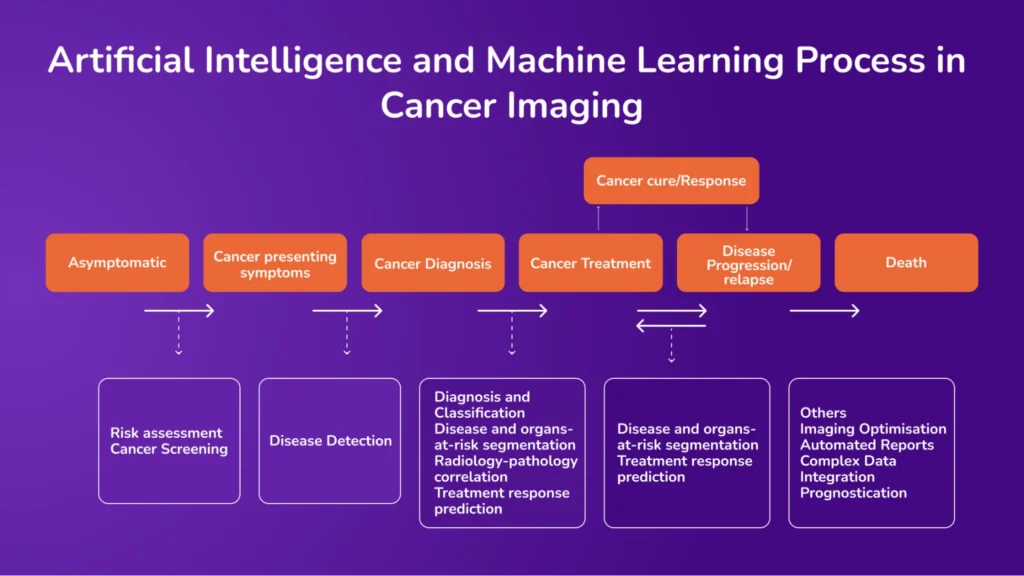
Applications of Medical Imaging Using Machine Learning and AI
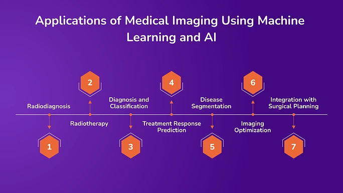
With the integration of artificial intelligence (AI), machine learning (ML), and medical image generation, medical imaging systems enable automated image analysis, reduce human error, and support clinical decision-making.
1. Radiodiagnosis
Modern imaging systems, such as computed tomography (CT) and magnetic resonance imaging (MRI), are incorporating machine learning (ML) techniques to enhance image acquisition and interpretation. CT scans provide detailed cross-sectional images of the body, which help assess the size and location of tumors. Advanced CT scanners can now capture full-body scans in high resolution within seconds.
MRI, known for its ability to distinguish soft tissues, is especially valuable in identifying cancers in the brain, joints, and internal organs. ML models, including neural networks and deep learning algorithms, support tasks such as brain tumor segmentation by distinguishing between cancerous and healthy tissue.
In breast cancer diagnosis, ultrasonography remains a vital imaging method. However, manually segmenting breast tissue is time-consuming and requires expertise. ML, particularly convolutional neural networks (CNNs), has been used to automate this process, effectively classifying tissues such as fatty, fibroglandular, and tumor areas in 3D ultrasound images. This automation improves accuracy and saves time in clinical workflows.
2. Radiotherapy
AI plays a vital role in radiotherapy by enhancing precision in treatment planning. Algorithms are used to define tumor boundaries accurately, ensuring that radiation targets the cancerous tissues while minimizing exposure to healthy structures.
AI also helps optimize treatment plans by analyzing large datasets, and it enables real-time monitoring of patient response, allowing timely adjustments to therapy. These capabilities improve treatment outcomes and reduce side effects.
3. Diagnosis and Classification
AI and ML models are applied to classify imaging findings and support diagnosis. In brain tumors, for example, ML systems can differentiate between various tumor types and grades based on imaging features, reducing the need for invasive biopsy procedures.
Similarly, AI is being used to classify cystic lesions in the pancreas and distinguish between benign and malignant growths with greater precision.
4. Treatment Response Prediction
Machine learning, combined with radiomics, is used to predict how tumors will respond to specific treatments. By analyzing pre-treatment images and extracting features such as shape, texture, and density, machine learning (ML) models can predict treatment outcomes. These predictions support personalized treatment planning and help avoid ineffective therapies.
5. Disease Segmentation
Segmentation of tumors and organs is essential for diagnosis, treatment planning, and monitoring disease progression. Deep learning models are capable of automating segmentation, improving consistency, and reducing the time required for manual outlining.
These models are particularly effective when applied to large datasets and high-resolution images, such as CT and MRI scans.
6. Imaging Optimization
AI is also used to improve imaging processes. For example, in MRI, machine learning (ML) algorithms are used to reduce scan times and enhance the quality of images. This helps reduce patient wait times and increases the efficiency of radiology departments. Super-resolution techniques enable more precise imaging, even when using faster, lower-resolution scans.
7. Integration with Surgical Planning
In surgical oncology, AI and computer vision technologies are being integrated into image-guided navigation systems. These tools use preoperative imaging to help surgeons identify vital anatomical structures and plan precise interventions. Such systems are increasingly used in neurosurgery, liver cancer surgery, and other areas where precision is critical.
As we continue to harness the power of medical image generation, it is clear that AI is set to play an even more critical role in transforming cancer research and treatment, ultimately improving patient care and survival rates.
Transforming Healthcare with Avahi AI: Streamlining Processes and Improving Care
The Avahi AI platform is playing a pivotal role in healthcare by providing innovative solutions that streamline medical data handling and enhance the quality of healthcare services. The platform offers several features to improve efficiency and accuracy in various aspects of healthcare, from document processing to medical transcription.
1. Smart PDF Summarizer
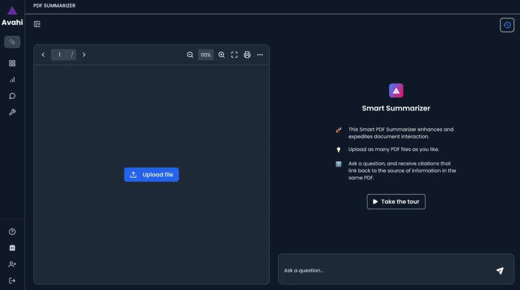
Avahi AI’s Smart PDF Summarizer makes it easier for healthcare professionals to interact with large documents, such as medical reports, research papers, and patient records. By uploading multiple PDF files, users can generate concise summaries while retaining key information.
This helps reduce the time spent on reviewing lengthy documents, enabling healthcare providers to access relevant insights for better decision-making quickly. Additionally, the platform allows you to view the original text alongside the summary, providing a more comprehensive understanding of the document’s content.
Benefits:
- Time-saving: Expedites the process of reviewing and summarizing large medical documents.
- Increased accuracy: Extracts key information from complex documents, reducing human error.
- Improved decision-making: Quickly provides healthcare professionals with actionable insights from medical records and research.
2. Data Masker
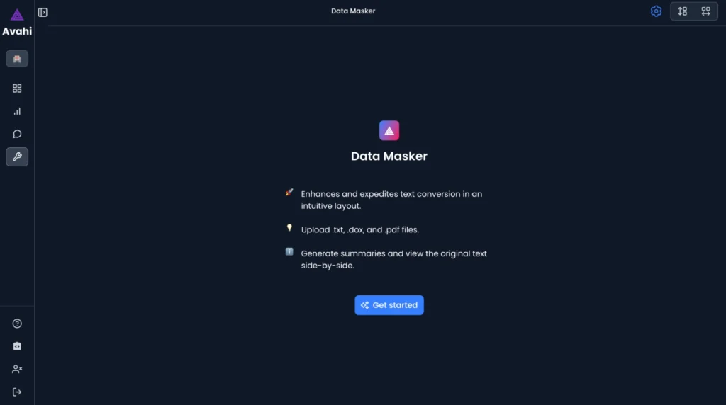
The Data Masker feature plays a crucial role in ensuring data privacy and security in healthcare settings. By masking sensitive patient data, this feature helps healthcare organizations comply with privacy regulations like HIPAA. This is especially beneficial when handling medical records or sharing data with third-party platforms, as it ensures that patient information is protected.
Benefits:
- Enhanced privacy: Protects sensitive patient information by anonymizing data.
- Regulatory compliance: Helps healthcare organizations meet legal requirements for data protection.
- Secure data sharing: Safely shares data with researchers or other healthcare providers without compromising patient confidentiality.
3. Medical Scribing

Avahi AI’s voice recording and medical scribing features support healthcare professionals by transcribing verbal notes into written text. This is particularly useful for physicians and medical staff who need to document patient interactions but don’t have much time to do so. By using voice recordings, healthcare providers can capture important patient information quickly and accurately without having to type out notes manually.
Benefits:
- Efficiency: Speeds up the documentation process by converting voice to text.
- Accuracy: Reduces the risk of errors that can occur when typing manually.
- Convenience: Allows healthcare providers to focus more on patient care while letting the AI platform handle the transcription.
Discover Avahi’s AI Platform in Action

At Avahi, we empower businesses to deploy advanced Generative AI that streamlines operations, enhances decision-making, and accelerates innovation—all with zero complexity.
As your trusted AWS Cloud Consulting Partner, we empower organizations to harness AI’s full potential while ensuring security, scalability, and compliance with industry-leading cloud solutions.
Our AI Solutions Include
- AI Adoption & Integration – Utilize Amazon Bedrock and GenAI to enhance automation and decision-making.
- Custom AI Development – Build intelligent applications tailored to your business needs.
- AI Model Optimization – Seamlessly switch between AI models with automated cost, accuracy, and performance comparisons.
- AI Automation – Automate repetitive tasks and free up time for strategic growth.
- Advanced Security & AI Governance – Ensure compliance, fraud detection, and secure model deployment.
Want to unlock the power of AI with enterprise-grade security and efficiency?
Get Started with Avahi’s AI Platform!
Frequently Asked Questions
- How is machine learning used to detect cancer earlier?
Machine learning models analyze patient records, medical images, and biomarker trends to spot early signs of cancer, often before symptoms appear. This supports proactive screenings and can improve survival outcomes by enabling earlier intervention.
- What is medical image generation, and why does it matter in cancer research?
Medical image generation uses AI (like GANs or diffusion models) to create synthetic but realistic scans. This helps researchers train models when real datasets are limited, especially for rare cancers. It boosts diagnostic accuracy while reducing data collection challenges.
- Can AI models outperform doctors in cancer diagnosis?
In some cases, yes. Deep learning models have matched or surpassed dermatologists in classifying skin lesions and are proving useful in areas like tumor segmentation and pathology. However, these tools are meant to support—not replace—clinical judgment.
- How does AI contribute to personalized cancer treatment?
AI helps predict how individual tumors respond to treatments like chemotherapy or immunotherapy. By analyzing tumor biology, genetics, and prior outcomes, machine learning enables more precise treatment plans, reducing trial-and-error in care.
- What makes Avahi AI useful for cancer research and care teams?
Avahi’s platform offers secure data masking, smart summarization of medical documents, and voice-based medical scribing—tools that save time, protect privacy, and streamline workflows. These features support faster research and better-informed clinical decisions.